andrew carnie
magic forest: slide dissolve work
The work arose from a collaboration
with Dr Richard Wingate, of the Medical Research Council Centre for Developmental
Neurology, at Kings College, London and was developed with the support of Dr Ken Arnold and Prof Marina Wallas. The work tracks the development,
proliferation, and organisation of neurones in the growing brain. The
work reflects 1. The changing organisation in the brain, developing to
being capable of holding memories, and 2. The process of collecting the
raw data for such scientific work through the use of the laser confocal
microscope.
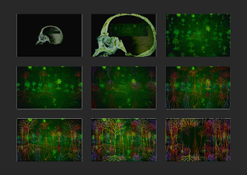
The work starts with the location of the growing brain in the skull and
proceeds with an ever-growing forest of neurones developing on the screens;
the mass increases, filling the whole screen with layers and layers of
neurones in different colours. The work ends when the system collapses
and the neurones disappear, blackness returns and the skull is shown again
getting larger and larger and the work begins to cycle around once more.
Each cycle lasts about fifteen minutes. The colours in the work reflect
the fluorescence used and seen in the staining of individual neurones,
which produce the images under the confocal microscope.
The brain consists of millions of neurons, each a finely
branching microscopic structure that reaches out for near and distant
neighbours; they contribute to a distributed circuitry whose logic remains
one of the greatest mysteries of biology. Structure and function are synonymous.
The geometry of the neuron determines its interconnections and is a physical
manifestation of the functional processing of the electrical signals it
receives. The complexity and beauty of its structure also reflects the
developmental constraints that shaped its growth.
Over a hundred years ago, the pioneer anatomist, Santiago Ramon y Cajal,
sliced up resin-impregnated brains which had been stained by a capricious
potassium dichromate-silver process invented by Camillo Golgi. For reasons
that are still not fully understood, a few cells in a thousand turn completely
black, their fine processes packed with dense particles.
By studying the fragments of cell distributed through the thin slices
of brain, individual neurons were imaginatively reconstructed and giving
rise to a model of the cellular composition of different brain regions,
their interconnections and even the direction of the flow of information.
Prior to Golgi's stain the very existence of cells in the brain was hotly
debated. Contrary evidence suggested that the brain was a continuous mesh
of interconnected fibres. A theory that was eventually superseded, as
the fine structure of neurons was uncovered in the early part of the twentieth
century.
Today, our understanding of the brain still requires the imaginative and
computer-aided analysis of slices through brain tissue. By using fluorescent
dyes, slices no longer have to be collected using a sharpened blade. Laser
scanning confocal microscopes can capture sections of stained neurons
optically, giving unprecedented images of the three-dimensional living
brain cells retained within computer memory. Just as our concepts
of neuroantaomy rely on sections of space, our ideas of how neurons grow
come from snapshots in time. Neurons are born within the inner lining
of the ventricular cavities that lie at the centre of the brain. They
migrate into outer layers of neurons forming layers and nuclei populations
of cells serving a particular function. They extend fine fibres, reaching
for appropriate neighbours to communicate with. One of these, the axon,
contributes to information highways connecting distant regions of the
brain. Finally, connections and fibres are remodelled as information itself
shapes the structure of individual cells. Piecing together brain cell
structure at different time-points has begun to give clues as to how cells
might interact and shape themselves as they grow. However our model of
brain development is still derived from glimpses of individual cells or
by identifying populations by means which inevitably obscure the fine
details of individual cells. Sections and snapshots remain, for the time
being, the basis of our understanding of neuroanatomy.
Richard Wingate, King's College, London
magic forest has been exhibited in:
the rotterdam natural history museum
the science museum london
the rotterdam film festival
the westport artcentre usa
the design museum zurich switzerland
british assocciation science festival in exeter
the art history dept oxford with martin kemp
the art and mind festival winchester
exit art new york
williams college museum of art
key of life leiden
to be exhibited next
'Magic Forest' will be shown again in the perra museum in istanbul in april 2011 and in dresden and Brno in 2011/2012
magic forest in art in america
| about us | site map | privacy policy | finding the studio | catalogue | contact us | finding city road winchester |
© 2004 Carnie Art Services